Search for the defocus value
Sample: single EM of K2O.7Nb2O5 at 1.9Å resolution
without ED data
Starting from
an average image (the left sub-window), by clicking the button 'Search
for
Df'  and following the direction of a few dialog boxes, a new sub-window (on
the right) will appear, which contains curves describing the variation
of figures of merit with different trial defocus values. The ordinate indicates
the value of figures of merit, while the abscissa indicates the value of
defocus.
and following the direction of a few dialog boxes, a new sub-window (on
the right) will appear, which contains curves describing the variation
of figures of merit with different trial defocus values. The ordinate indicates
the value of figures of merit, while the abscissa indicates the value of
defocus.
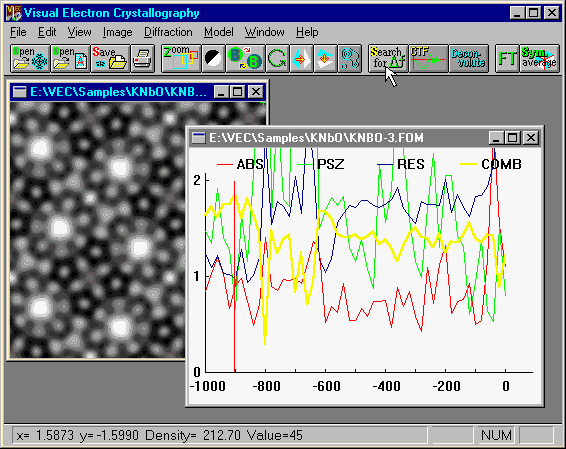
In the plot of figures of merit, the red curve denotes the
absolute figure of merit, blue denotes the
residual
figure of merit, green denotes the psizero
figure of merit, while the yellow one denotes the combined
figure of merit, which is calculated from the previous three. The accepted
defocus value ( -900 Angstrom in this example) corresponds to the maximum
of the combined figure of merit.
Details of
the above figures of merit can be found in the book Physical and Non-physical
Methods of Solving Crystal Structurees by M.M. Woolfson and Fan Hai-fu,
Canbridge Univ. Press, 1995, pp.106-107.
For the theory
of finding the defocus value from a single electron micrograph, the user
is referred to Han, F.S., Fan, H.F. and Li, F.H. (1986). Acta Cryst., A42,353-6.
