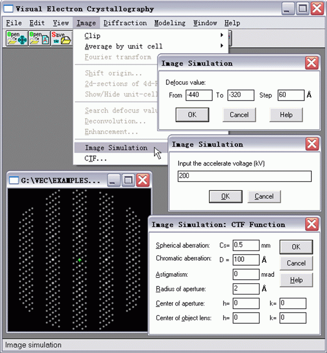
Starting from a single retrieved dynamical electron diffraction pattern (see the bottom-left sub-window on the figure below), click the pulldown menu Diffraction and select the iterm Image Simulation.

Fill necessary values into the dialog boxes and click OK. Then VEC will calculate and store the resultant EMs as *.ave files. A messege like the following will finally appear.

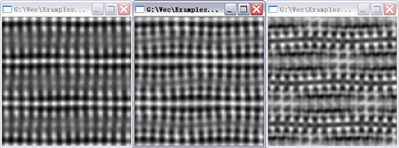
To display the simulated EMs, the user should open them in VEC in graphic mode (shown below). Defocus values of the following EMs are from left to right respectively -320 Å, -380 Å and -440 Å. Other parameters can be found in the last two dialog boxes of the first figure.

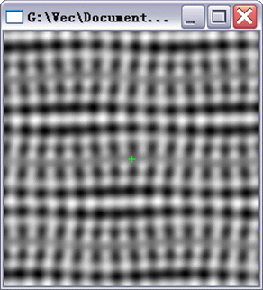
For comparison, the experimental EM of Bi-2201 is shown below, which is taken near Scherzer focus with a JEOL 2010 high-resolution electron microscope under the condition: accelerating voltage U = 200 kV, spherical aberration Cs = 0.5 mm, defocus spread D = 70 Å, defocus value Δf = -380 Å and divergence half-angle α = 0.01 mrad.
